Programmed death-1 (PD-1) is a member of the CD28 family of receptors that includes CD28, cytotoxic T-lymphocyte-associa
ted antigen 4 (CTLA-4), inducible costimulator(ICOS), and Band T-lymphocyte attenuator. These receptors play a role in the cellularimmune response. For example, CD28 serves as a costimulatory receptor that enhances T-cell activation, whereas CTLA-4 serves as an inhibitor of T-cell activation. PD-1 also hasan inhibitory function on T cells and B cells, and is important in peripheral tolerance. There are at least 2 ligands for PD-1: PD-L1 and PD-L2, which are expressed on a range of cells. CD28 is constitutively expressed on most or all CD4+ T cells and approximately 50% of CD8+ T cells, whereas CTLA-4 is not expressed on resting T cells. PD-1 is also expressed on activated T cells, B cells, and myeloid cells. Iwai and coworkers studied the microanatomic distribution of PD-1 in human tonsil and found that PD-1 is expressed on most T cells and a small subset of B cells in the light zone of germinal centers, but not elsewhere in the tonsil. On that basis, it was postulated that PD-1 may play a role in the process of clonal selection of centrocytes, which occurs in this subanatomic site in germinal centers. PD-1 is a new marker of angioimmunoblastic lymphoma and suggests a unique cell of origin for this neoplasm. Unlike CD10 and bcl-6, PD-1 is expressed by few B cells, so it maybe a more specific and useful diagnostic marker in angioimmunoblastic lymphoma. It also seems to stain a greater percentage of CD3-positive neoplastic cells in angioimmunoblastic lymphoma than either CD10 or bcl-6. In addition, PD-1 expression provides new evidence that angioimmunoblastic lymphoma is a neoplasm derived from germinal center-associated T cells. PD-1 expression in angioimmunoblastic lymphoma lends further support to this model of T-cell oncogenesis, in which specific subtypes of T cells may undergo neoplastic transformation and result in specific distinct histologic, immunophenotypic, and clinical subtypes of T-cell neoplasia.
Programmed death-1 (PD-1) is a member of the CD28 family of receptors that includes CD28, cytotoxic T-lymphocyte-associated antigen 4 (CTLA-4), inducible costimulator(ICOS), and Band T-lymphocyte attenuator. These receptors play a role in the cellularimmune response. For example, CD28 serves as a costimulatory receptor that enhances T-cell activation, whereas CTLA-4 serves as an inhibitor of T-cell activation. PD-1 also hasan inhibitory function on T cells and B cells, and is important in peripheral tolerance. There are at least 2 ligands for PD-1: PD-L1 and PD-L2, which are expressed on a range of cells.
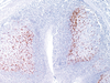
CD28 is constitutively expressed on most or all CD4+ T cells and approximately 50% of CD8+ T cells, whereas CTLA-4 is not expressed on resting T cells. PD-1 is also expressed on activated T cells, B cells, and myeloid cells. Iwai and coworkers studied the microanatomic distribution of PD-1 in human tonsil and found that PD-1 is expressed on most T cells and a small subset of B cells in the light zone of germinal centers, but not elsewhere in the tonsil. On that basis, it was postulated that PD-1 may play a role in the process of clonal selection of centrocytes, which occurs in this subanatomic site in germinal centers.
PD-1 is a new marker of angioimmunoblastic lymphoma and suggests a unique cell of origin for this neoplasm. Unlike CD10 and bcl-6, PD-1 is expressed by few B cells, so it maybe a more specific and useful diagnostic marker in angioimmunoblastic lymphoma. It also seems to stain a greater percentage of CD3-positive neoplastic cells in angioimmunoblastic lymphoma than either CD10 or bcl-6. In addition, PD-1 expression provides new evidence that angioimmunoblastic lymphoma is a neoplasm derived from germinal center-associated T cells. PD-1 expression in angioimmunoblastic lymphoma lends further support to this model of T-cell oncogenesis, in which specific subtypes of T cells may undergo neoplastic transformation and result in specific distinct histologic, immunophenotypic, and clinical subtypes of T-cell neoplasia.