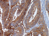

Ep-CAM consists of two glycoproteins, 34 and 39 kDa, sometimes designated epithelial antigen, epithelial specific antigen, and epithelial glycoprotein. The glycoproteins are located on the cell membrane surface and in the cytoplasm of virtually all epithelial cells with the exception of most squamous epithelia, hepatocytes, renal proximal tubular cells, gastric parietal cells and myoepithelial cells. However, focal positivity may be seen in the basal layer of squamous cell epithelium of endoderm (e.g., palatine tonsils) and mesoderm (e.g., uterine cervix). In liver lesions like hepatitis and cirrhosis, the hepatocytes frequently become Ep-CAM positive. Normal mesothelial cells are Ep-CAM negative, but may express focal reaction when undergoing reactive changes. Mesenchymal cells and cells from the neural crest are negative, with the exception of olfactory neurons.
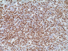
Ep-CAM is found in the large majority of adenocarcinomas of most sites (50-100% invarious studies); as well as neuroendocrine tumours, including small cell carcinoma. Renal cell carcinoma and hepatocellular carcinoma stain in about 30% of the cases. Squamous cell carcinoma of endoderm and mesoderm is usually Ber-EP4 positive, while that of ectoderm is negative. Basal cell and basosquamous carcinoma are Ber-EP4 positive in almost all cases. Choroid plexus papilloma and carcinoma are usually negative. Malignant mesothelioma (epithelioid and biphasic) is Ep-CAM positive in 4-26% of the cases. The staining is usually focal, but may occasionally be widespread. Synovial sarcoma (epitheloid and biphasic) and desmoplastic small round cell tumour stain for Ep-CAM in most cases.
Seminoma, embryonal carcinoma, yolk sac tumor and choriocarcinoma reveal Ber-EP4 positivity in a minor proportion of cases. Among neural tumours, Ep-CAM positivity is seen only in olfactory neuroblastoma. Ep-CAM can be of great help in the differential diagnosis of malignant involvement in the peritoneal and pleural cavities. The lack of reactivity in the majority of malignant mesothelioma can in an appropriate panel be utilized in the discrimination between this tumour and adenocarcinoma.
| Ventana Medical Systems, Inc. | |
|---|---|
| Product Category | Organic Chemicals |
| Product Number | 760-4383 |
| Product Name | Ep-CAM (Epithelial Specific Antigen) |